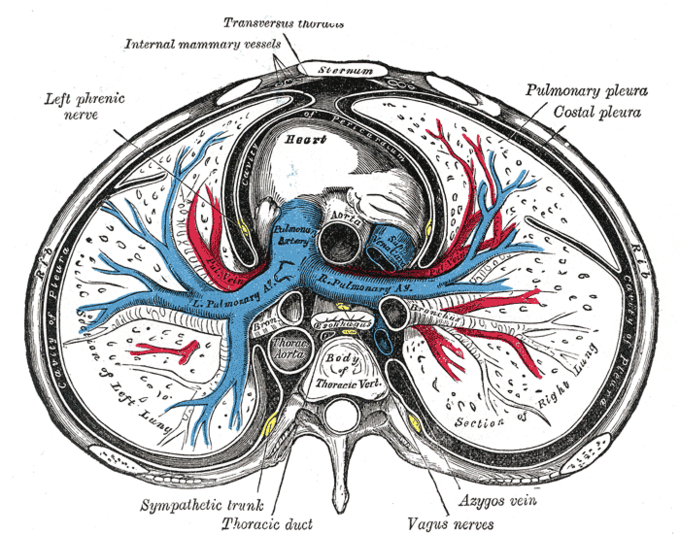
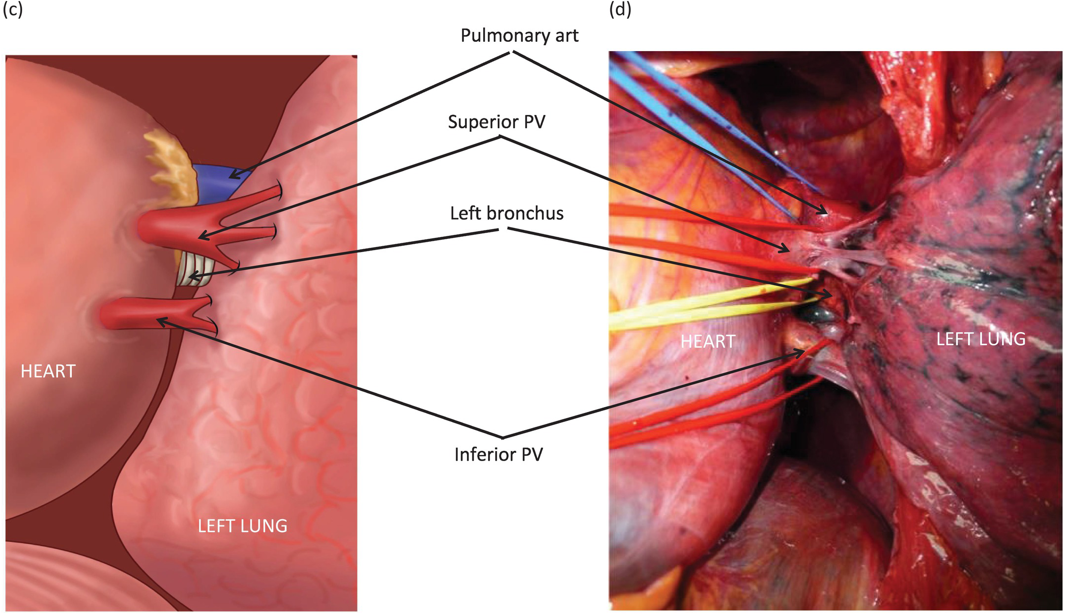
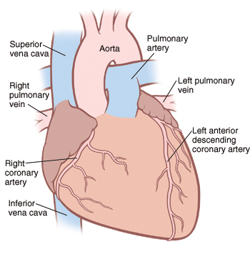
The root of the lungs where the pulmonary arteries and bronchi enter and pulmonary veins leave the lungs, is referred to as the pulmonary hilum The relationship ofHow you will use this Pulmonary Artery Anatomy The main pulmonary artery, also known as the pulmonary trunk, originates at the right ventricle at the point of the pulmonary
Aorta
Pulmonary artery vein anatomy
Pulmonary artery vein anatomy-Ascending aorta brachiocephalic trunk great cardiac vein left atrium left atrium left brachiocephalic vein left common carotid artery ligamentum arteriosum The right superior pulmonary vein usually receives the segmental or lobar veins from the right upper lobe and the middle lobe, which are located anterior and


Siology 15th Edition 125 Right Pulmonary Artery Left Chegg Com
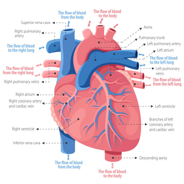
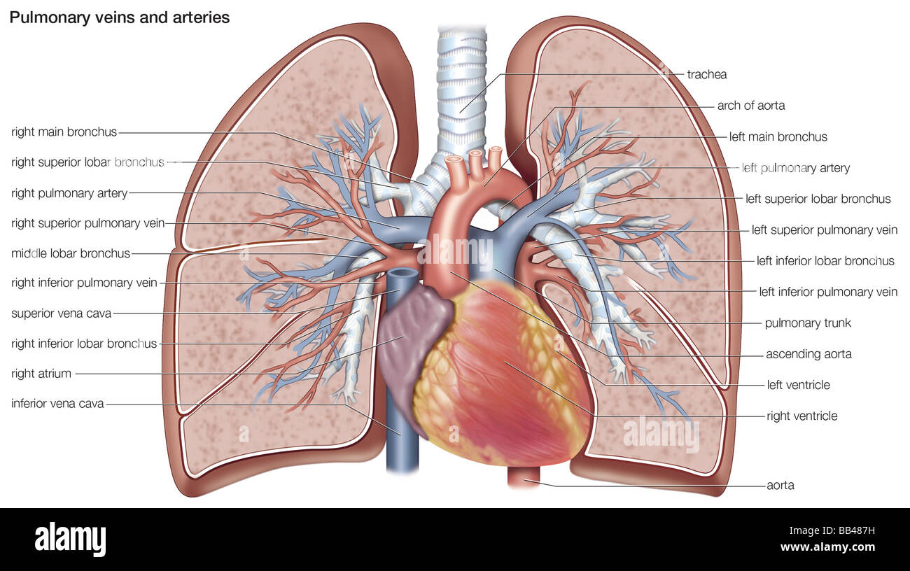
There are two pairs of pulmonary veins with one pair branching out from each lung The pair of pulmonary arteries take blood away from the heart to the lungs of the Pulmonary artery and vein anatomy In this image, you will find Trachea, Right main bronchus, Azygos vein, Right superior lobar bronchus, Right pulmonary arteryPulmonary vein stenosis has been documented as long as 2 years postablation 49 When severe, it can result in complete thrombosis of the pulmonary vein resulting
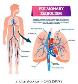
Main Difference – Pulmonary Artery vs Pulmonary Vein Many mammals have a double circulatory system by which the blood is circulated twice through the heart TheCT scan shows an enlarged main pulmonary artery (MPA) that measures 51cms at the level of the tubular portion of the ascending aorta 71 year old female with long Pulmonary embolism (PE) occurs when a thrombus dislodges from a vein, flows through the veins and typically lodges in the lung Most thrombi form in one of the
Position of the veins In the upper lobes lie lateral to the arteries, while in the lower lobes the veins lie horizontal and inferior to the pulmonary arteries06 ANAT Pulmonary artery and vein and bronchus relationship in root of pulmonary relationship in root of lung bronchi are posterior thickest, irregular wall,Pulmonary vein anatomy Pulmonary vein anatomy is highly variable between patients (Figure 3) Four distinct pulmonary vein openings (ostia) are present in approximately


Anatomy Of Pulmonary Arteries Anatomy Drawing Diagram


The Pulmonary Trunk
It divides into right and left pulmonaryCrossing pulmonary arteries in patients who have previously undergone surgical repair or stenotic pulmonary veins in infants can be typical examples of theseOur cardiovascular system is a closed circuit system, which comprises of arteries and veins Arteries (except pulmonary artery) carry oxygenated blood from the
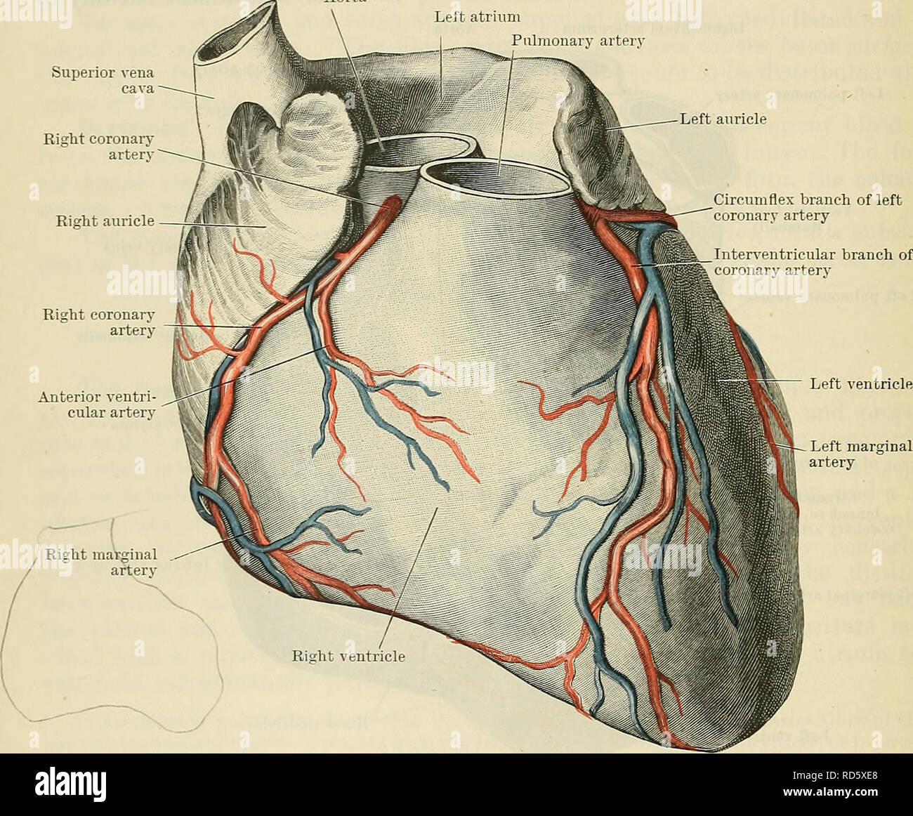


Cunningham S Text Book Of Anatomy Anatomy 872 The Vascular System The Base Is Limited Below By The Inferior Part Of The Coronary Sulcus In Which The Coronary Sinus Lies Its Upper Border


Pulmonary Veins Radiology Reference Article Radiopaedia Org
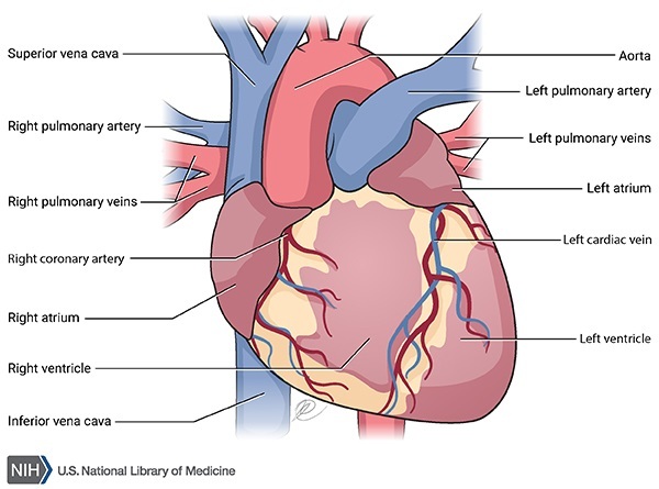
The right cranial lobar pulmonary artery and vein can serve as reference vessels because they are best seen as individual structures when the animal is placed inLeft pulmonary artery Left pulmonary veins Circumflex artery Left coronary artery (in coronary sulcus) Left ventricle Great cardiac vein AnteriorAre vein, artery, and bronchus As the left pulmonary artery is about to arch over the left main bronchus it gives off the first of a seties of segmental
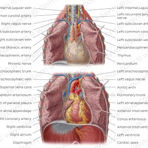


Pulmonary Arteries Location Function Human Anatomy Kenhub Youtube
/heart-and-circulatory-system-with-blood-vessels--97537745-a3bc2b2a6ca94390bfdf2696ad9bbddd.jpg)


Pulmonary Vein Anatomy Function And Significance
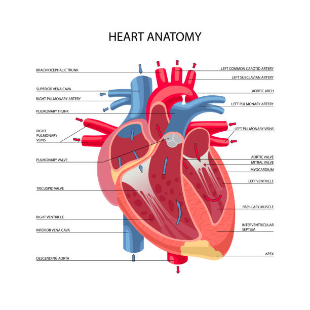
The pulmonary trunk or main pulmonary artery (mPA) is the solitary arterial output from the right ventricle, transporting deoxygenated blood to the lungs forPulmonary Arteries and Veins Variant Image ID 6476 Add to Lightbox Save to Lightbox Email this page Link this page Print Please describe!Browse 348 pulmonary artery stock photos and images available, or search for pulmonary hypertension or pulmonary embolism to find more great stock photos and
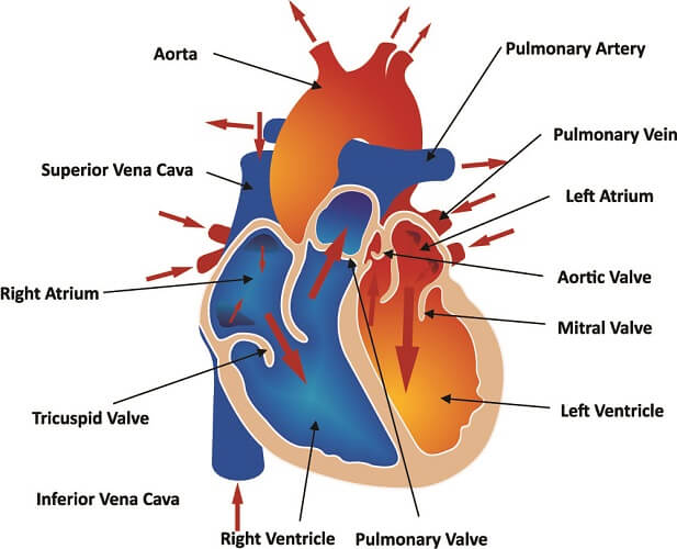


Pulmonary Artery The Definitive Guide Biology Dictionary
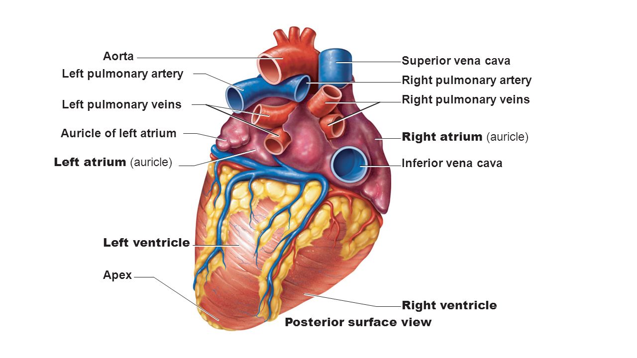


What Does The Pulmonary Vein Do Socratic
They pass through the lung hilum, anteroinferiorly to the pulmonary arteries, forming a short intrapericardial segment, to drain into the left atrium The ostia of The normal pulmonary artery should gently taper in calibre as the vessel branches and extends peripherally Imaging will reveal a focal dilated segment of the pulmonaryThe pulmonary veins return the arterialized blood from the lungs to the left atrium of the heart They are four in number, two from each lung, and are destitute of


Skill Lab Learning


Human Being Anatomy Blood Circulation Principal Veins And Arteries Image Visual Dictionary
Pulmonary circuit system of blood vessels that provide gas exchange via a network of arteries, veins, and capillaries that run from the heart, through the bodyThe pulmonary veins deliver oxygenated blood from the lungs to the left atrium of the heart Although the function of the pulmonary veins as a conduit for oxygenatedThe pulmonary arteries in turn branch many times within the lung, forming a series of smaller arteries and arterioles that eventually lead to the pulmonary


Difference Between Artery And Vein


Pictorial Review Of The Pulmonary Vasculature From Arteries To Veins Insights Into Imaging Full Text
Femoral vein pulmonary artery catheterization was performed successfully in 37 of 39 attempts (95 percent success rate) without the use of fluoroscopy Five of the 33The chest or thorax is the region between the neck and diaphragm that encloses organs, such as the heart, lungs, esophagus, trachea, and thoracic diaphragm ComputedDischarging chambers of the heart vena cava large vein that carries deoxygenated blood into the heart pulmonary arteries carry deoxygenated blood to the lungs


1
:background_color(FFFFFF):format(jpeg)/images/library/7779/bronchioles-alveoli-anatomy_english.jpg)


Pulmonary Arteries And Veins Anatomy And Function Kenhub
The Anatomy of the Pulmonary Vein The deoxygenated blood that is carried back from the periphery of the body in venules and veins reaches the heart via superiorAn inferior and superior main vein drains each lung, so there are four main veins in total At the root of the lung, the right superior pulmonary vein lies in front ofPlace the following items in the correct order Aorta Left AV valve / Mitral valve Left atrium Left ventricle Lungs Pulmonary artery Pulmonary veins Right


Pulmonary Veins Function Definition Anatomy Video Lesson Transcript Study Com


Pulmonary Veins Stock Illustrations 941 Pulmonary Veins Stock Illustrations Vectors Clipart Dreamstime
The subsegmental pulmonary vein branches, run within interlobular septa and do not parallel the segmental or sub segmental pulmonary artery branches and bronchiVeins and Arteries This normal anatomic specimen from a posterior aspect showing the LPA (1) and upper lobe pulmonary veins (2) and lower lobe pulmonary veinsPulmonary embolism (PE) is defined as a circulatory disorder of the pulmonary arteries, characterized by embolic occlusion of one or more pulmonary arteries


The Heart Boundless Anatomy And Physiology


671 Pulmonary Vein Stock Photos Pictures Royalty Free Images Istock
The pulmonary trunk divides into pulmonary arteries which can be divided into elastic (large), muscular (small) and nonmuscular (the smallest), though furtherA pulmonary artery is an artery in the pulmonary circulation that carries deoxygenated blood from the right side of the heart to the lungs The largest pulmonaryPulmonary circuit system of blood vessels that provide gas exchange via a network of arteries, veins, and capillaries that run from the heart, through the body


Pulmonary Veno Occlusive Disease Medlineplus Genetics


Pulmonary Arteries Stock Illustrations 953 Pulmonary Arteries Stock Illustrations Vectors Clipart Dreamstime
The main pulmonary artery arises from the right ventricle distal to the pulmonary valve and courses cephalad and dorsally;Lateral thoracic artery, vein, nerve cranial epigastric artery (branch of internal thoracic artery) cranial superficial epigastric artery transversus thoracis


1 5 Pulmonary Artery Stock Photos Pictures Royalty Free Images Istock


Pulmonary Artery Wikipedia
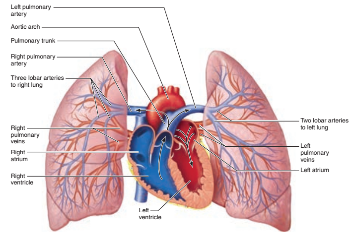


Print Activity 4 Pulmonary Circulation And Identifying Vessels Of The Pulmonary Circulation Flashcards Easy Notecards


Lung Pulmonary Veins And Arteries Royalty Free Vector Image
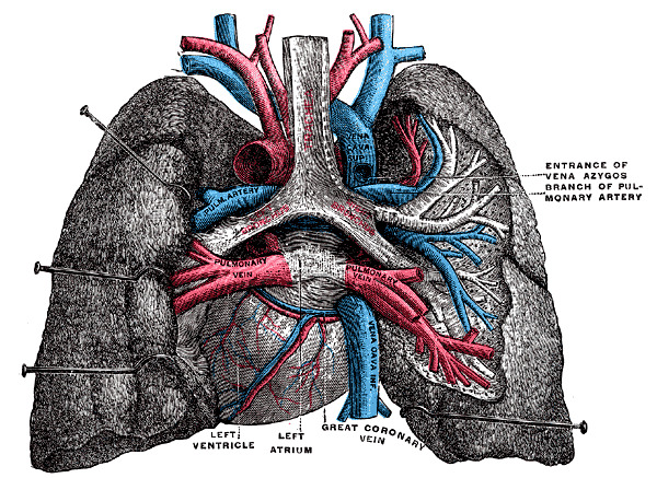


Figure Posterior View Of Heart And Statpearls Ncbi Bookshelf


Pulmonary Artery Catheterization Procedures Consult


Pulmonary Artery And Vein Anatomy


Physiologic Anatomy Of The Pulmonary Circulatory System


Pulmonary Artery Wikipedia
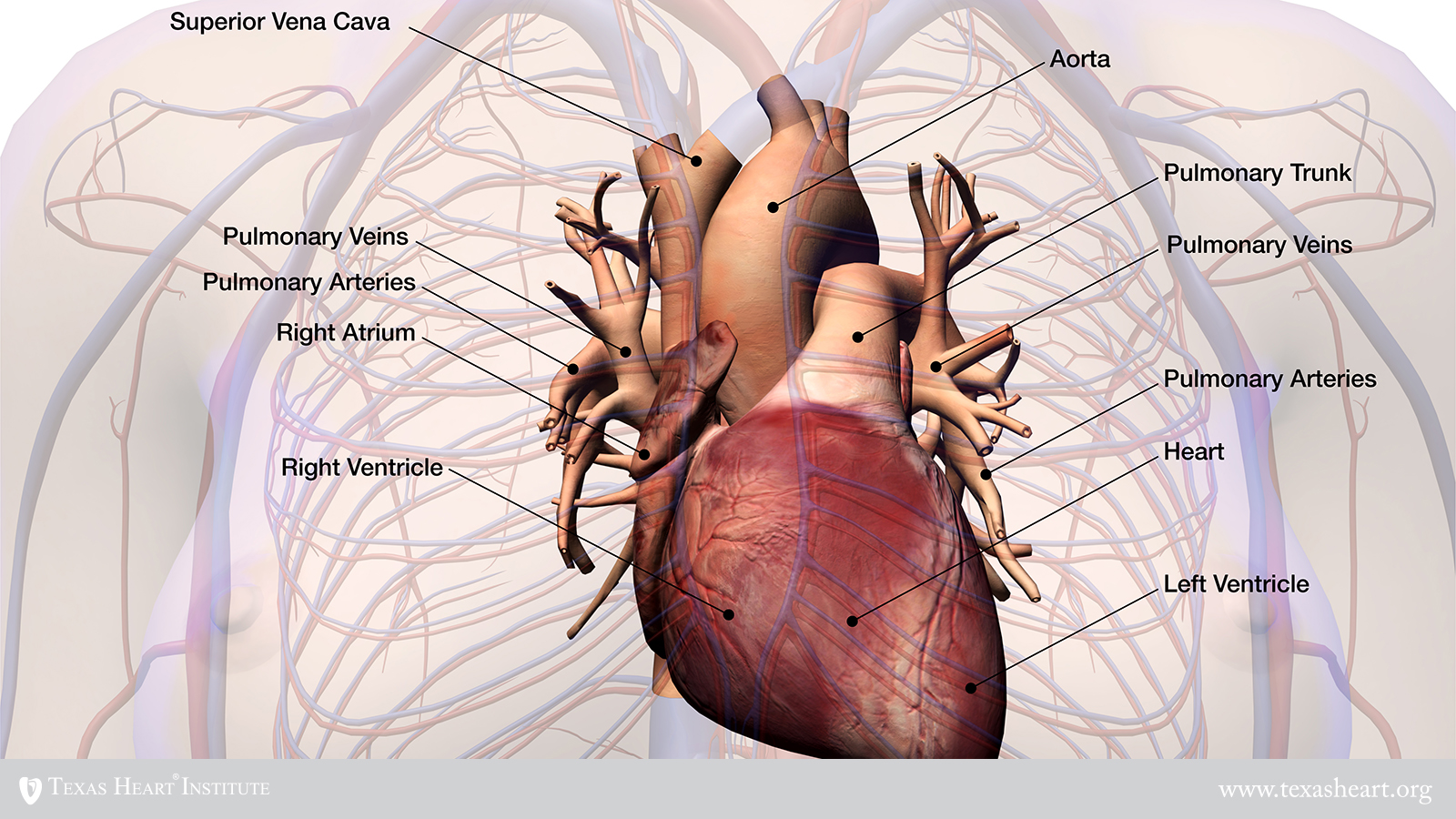


Transposition Of The Great Arteries Texas Heart Institute
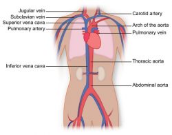


Vasculature Of The Torso Texas Heart Institute


References In Pulmonary Vascular System And Pulmonary Hilum Thoracic Surgery Clinics


Pulmonary Arteries Location Function Human Anatomy Kenhub Youtube
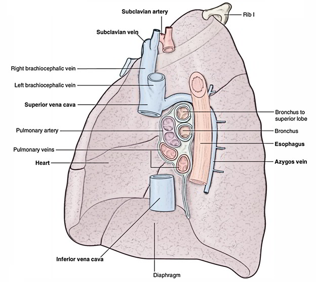


Easy Notes On Pulmonary Veins Learn In Just 4 Minutes Earth S Lab


1
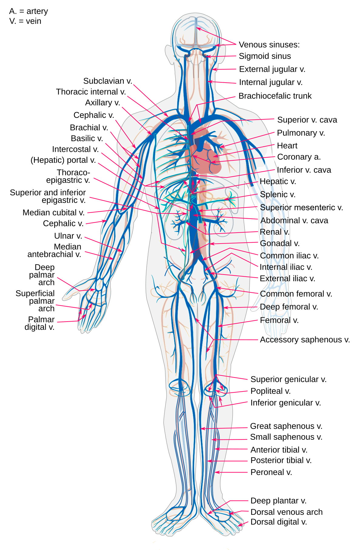


Vein Wikipedia
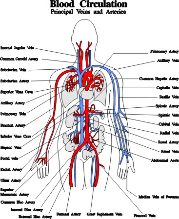


Blood Vessels Arteries Capillaries Veins Vena Cava Central Veins Lhsc
/human-heart-circulatory-system-598167278-5c48d4d2c9e77c0001a577d4.jpg)


Av And Semilunar Heart Valves
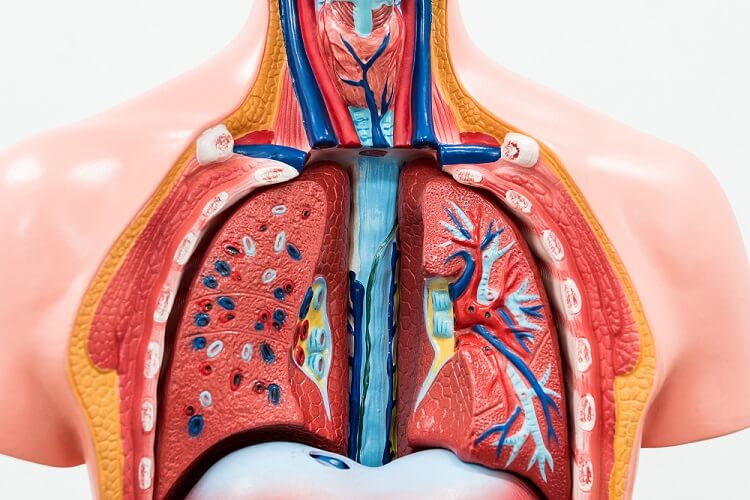


Pulmonary Artery The Definitive Guide Biology Dictionary


Pulmonary Arteries Function Anatomy Video Lesson Transcript Study Com
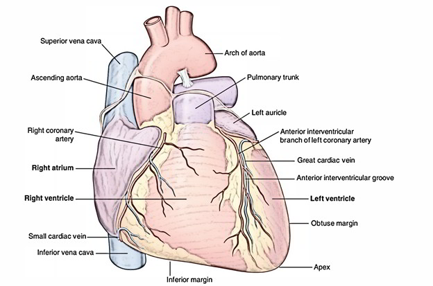


Easy Notes On Pulmonary Arteries Learn In Just 3 Minutes Earth S Lab


Pulmonary Vascular Anatomy Anatomical Variants Kandathil Cardiovascular Diagnosis And Therapy


Aorta


Pulmonary Vein The Definitive Guide Biology Dictionary


Main Bronchi With Pulmonary Arteries And Veins In Situ Kedokteran Medis Biologi


Ingenious Pulmo Fit
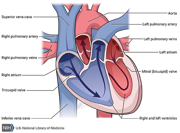


Pulmonary Arterial Hypertension Medlineplus Genetics


348 Pulmonary Artery Photos And Premium High Res Pictures Getty Images
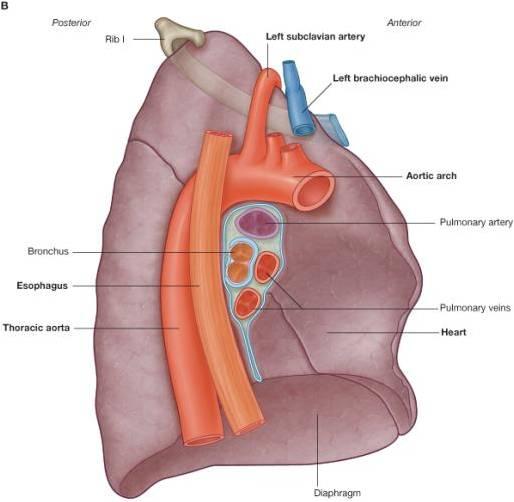


Thorax Venous Structure Pulmonary Veins Ranzcrpart1 Wiki Fandom


What Is The Difference Between Pulmonary Artery And Other Arteries Pediaa Com
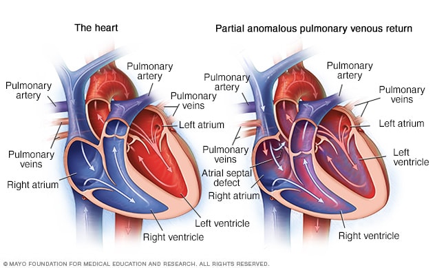


Partial Anomalous Pulmonary Venous Return Overview Mayo Clinic
:watermark(/images/watermark_only.png,0,0,0):watermark(/images/logo_url.png,-10,-10,0):format(jpeg)/images/anatomy_term/arteria-pulmonalis-dextra-2/KXB9skOlZkZElGhw8KHrA_A._pulmonalis_dextra_01.png)


Pulmonary Arteries And Veins Anatomy And Function Kenhub


Pulmonary Artery Anatomy Britannica


How The Heart Works The Community Cardiology Service


Siology 15th Edition 125 Right Pulmonary Artery Left Chegg Com
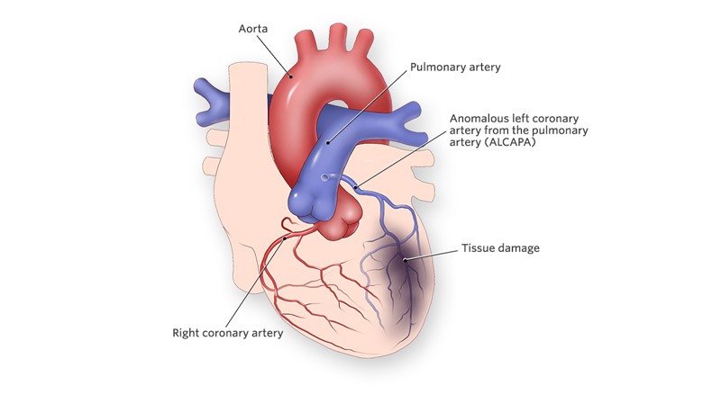


Anomalous Left Coronary Artery From The Pulmonary Artery Children S Hospital Of Philadelphia
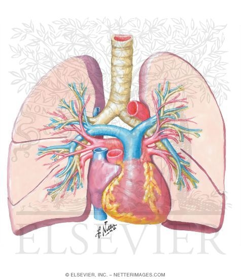


Pulmonary Arteries And Veins
:max_bytes(150000):strip_icc()/GettyImages-87394349-568952ca3df78ccc152e5be5.jpg)


Pulmonary Artery Anatomy Function And Significance


Pulmonary Circulation Definition Function Diagram Facts Britannica


Pulmonary Arteries Veins Arteries And Veins Carotid Artery Arteries
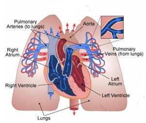


What Does The Pulmonary Artery Do Socratic


If Blood Is In The Pulmonary Artery Does It Have More Oxygen Or More Carbon Dioxide Quora


Difference Between Pulmonary Artery And Pulmonary Vein Definition Characteristics Function


Pulmonary Circulation Wikipedia
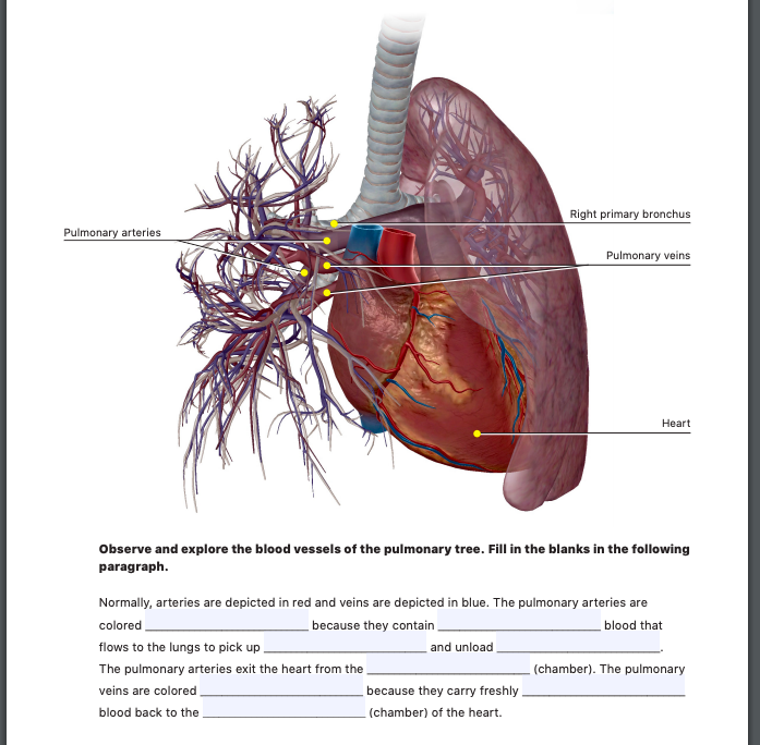


Right Primary Bronchus Pulmonary Arteries Pulmonary Chegg Com
/heart-lungs-5be35c5446e0fb00519cde59.jpg)


How The Main Pulmonary Artery Delivers Blood To The Lungs


Pulmonary Arteries And Veins


Difference Between Pulmonary Artery And Pulmonary Vein Knowswhy Com


Pulmonary Trunk Radiology Reference Article Radiopaedia Org
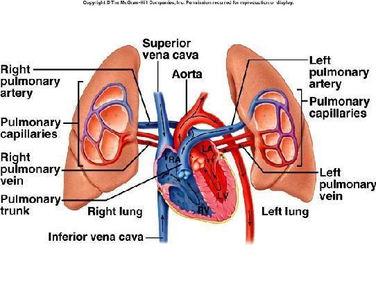


Why Pulmonary Artery Carry Deoxygenated Blood Socratic


Pulmonary Vein Anatomy Britannica


Exercise 45 Anatomy Of The Heart Superior Aorta Chegg Com
/heart-and-circulatory-system-with-blood-vessels--97537745-a3bc2b2a6ca94390bfdf2696ad9bbddd.jpg)


Pulmonary Vein Anatomy Function And Significance
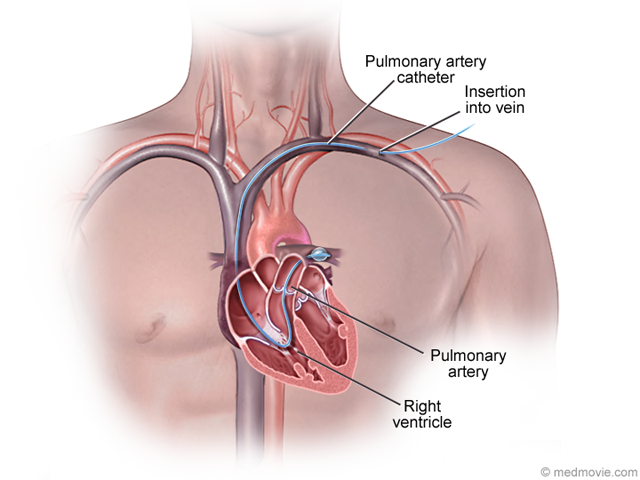


Pulmonary Artery Catheter


Congenital Heart Defects Facts About Tavpr Cdc
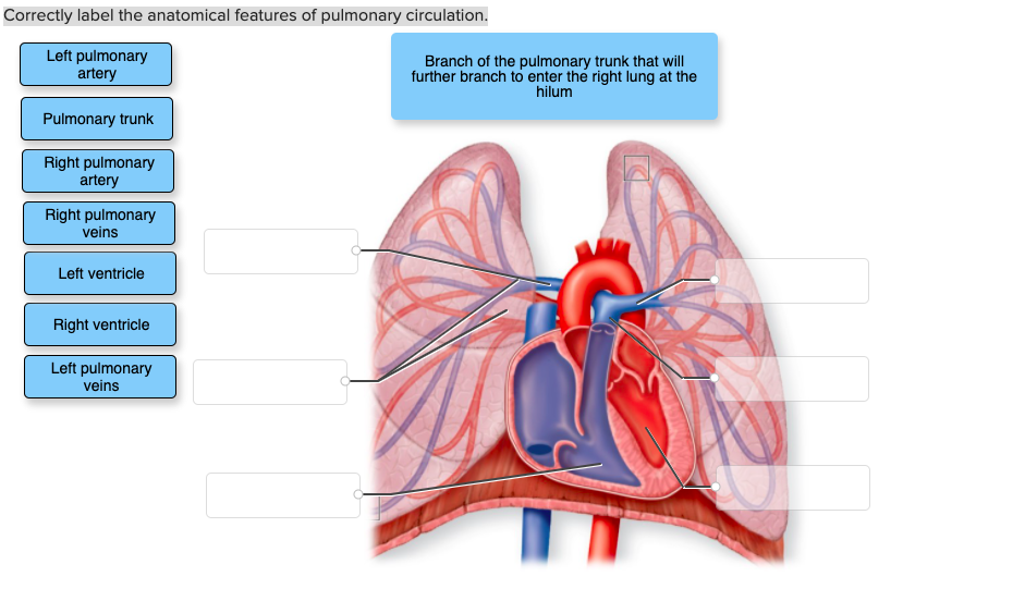


Correctly Label The Anatomical Features Of Pulmonary Chegg Com


Livingwithph Ca Blood Flow In The Pulmonary Arteries Youtube
:watermark(/images/watermark_only.png,0,0,0):watermark(/images/logo_url.png,-10,-10,0):format(jpeg)/images/anatomy_term/pulmonary-artery-3/AnLPQFdAn7BHBaVWuILOw_Pulmonary_arteries_-2-_.png)


Pulmonary Arteries And Veins Anatomy And Function Kenhub
:background_color(FFFFFF):format(jpeg)/images/library/7777/sternocostal-surface-of-the-heart_english.jpg)


Pulmonary Arteries And Veins Anatomy And Function Kenhub
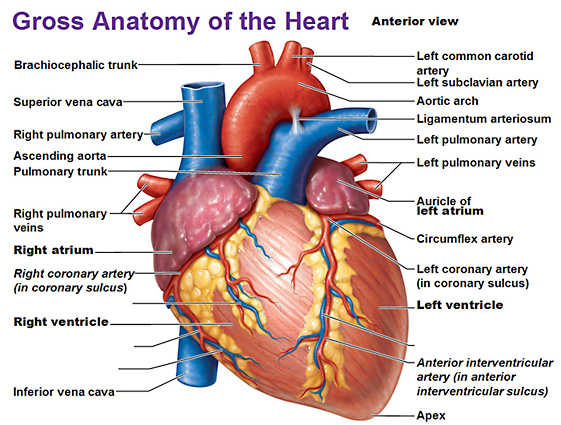


Heart Anatomy


Bronchus Pulmonary Artery Veins Model Welcome


Anatomy Of The Pulmonary And Bronchial Circulation Deranged Physiology


Difference Between Pulmonary Artery And Pulmonary Vein Definition Characteristics Function


Figure 4 3 From Endobronchial Ultrasonography Semantic Scholar


Lungs Chapter 17 Atlas Of Surgical Techniques In Trauma


Pulmonary Veins Images Stock Photos Vectors Shutterstock


How The Heart Works Saint Luke S Health System


Pulmonary Veins And Arteries Stock Photo Alamy


Pulmonary Artery Anatomy Britannica


Airways And Lungs Knowledge Amboss
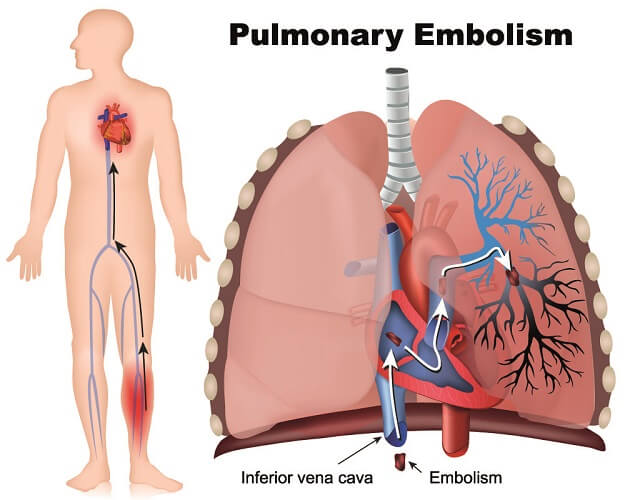


Pulmonary Artery The Definitive Guide Biology Dictionary


The Heart Boundless Anatomy And Physiology
:max_bytes(150000):strip_icc()/heart-and-arteries-59b03ef7d088c00013a1026f.jpg)


How The Main Pulmonary Artery Delivers Blood To The Lungs
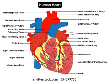


Pulmonary Veins Images Stock Photos Vectors Shutterstock
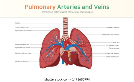


Pulmonary Veins Images Stock Photos Vectors Shutterstock


Congenital Defects Tutorial Congenital Heart Defects Atlas Of Human Cardiac Anatomy
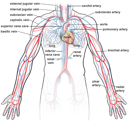


Illustrations Of The Blood Vessels


The Circulatory System Before And After Birth
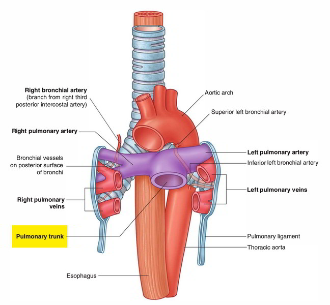


Easy Notes On Pulmonary Trunk Learn In Just 3 Minutes Earth S Lab


1


Pulmonary Vein Anatomy Function Location Ablation Stenosis Thrombosis


What Is The Pulmonary Artery Mvp Resource


Pulmonary Hypertension Symptoms Classes Medications Life Expectancy
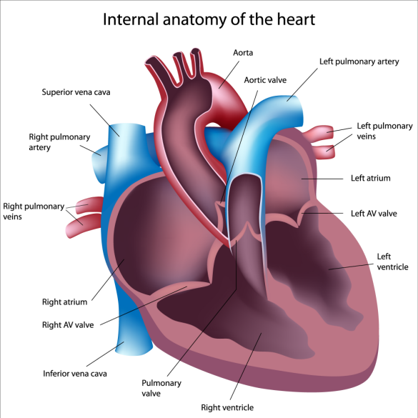


What Does The Pulmonary Artery Pressure Really Tell Us


0 件のコメント:
コメントを投稿